
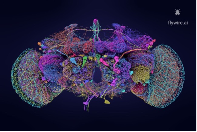


As neuroscientists’ interest in brain functionality and mental disorders has grown, so too has the demand for more precise neural mapping techniques. With more than 80 billion neurons and trillion connections, the creation of a “connectome” – or wiring diagram – for the human brain remains an unfathomably complex task. This complexity has, until recently, hindered researchers’ ability to link brain structure with function in ways that could drive targeted interventions for neuropsychiatric disorders or neurodegenerative diseases. However, with initiatives like the FlyWire project at the forefront of high-resolution neural mapping, a human brain connectome is becoming increasingly achievable and may soon be the nexus of many neuroscientific breakthroughs.
Early this October, FlyWire – a global consortium comprising dozens of research labs – published a nine-paper package in the Nature Journal,detailing its success in producing a fully annotated neuronal wiring diagram for the adult fruit fly, Drosophila melanogaster. While complete connectomes for a few, simpler organisms already existed, notably the roundworm Caenorhabditis elegans, their comparatively smaller brains offered limited applicability to human research. Thus, to create a structural foundation from which functional insights about the circuit dynamics and behaviour of our brains could arise, FlyWire researchers leveraged advanced electron-microscopy and computational technologies to reconstruct the Drosophila brain with unprecedented resolution.
Why the Drosophila brain and not that of other small organisms, you may ask? Well, with 139,255 neurons, the Drosophila offers an ideal balance of complexity and manageability. Though it contains a million times fewer neurons than that of humans, fruit flies can still exhibit a range of sophisticated behaviors such as navigation, learning, decision-making, and social interactions – all of which are high-order cognitive functions. The Drosophila emerged as a key model organism, in a large part due to the concentration of collaboratively generated molecular, genetic, and digital resources available for it. For instance, a partial fruit fly hemibrain connectome had already been proposed in 2020, and this provided a basis for FlyWire’s annotations.
The types of electron microscopy used in the reconstruction of this connectome were transmission electron microscopy (TEM) and scanning electron microscopy (SEM). TEM was applied by transmitting a high-energy electron beam through ultra-thin slices of brain tissue, allowing for the visualisation of intracellular structures, such as synapses and neuronal circuits. SEM was used to scan the surface of the tissue to produce detailed, three-dimensional images of the external morphology of neurons as well as enable the mapping of neuronal arrangements at a macrostructural level. The images captured by TEM and SEM were then processed and developed by the FlyWire team, who stitched them together and aligned them using advanced computational methods to create a cohesive digital model of Drosophila’s neural circuitry. A computational infrastructure was also used to proofread the neuron reconstructions – an effort that would have taken 33 years had it been done by a single person working full time.

However, a connectome is only useful if researchers can readily identify neurons of interest, and so the annotation process began. Once the basic metadata for each neuron – such as soma position, laterality, and the corresponding afferent or efferent nerve connections – had been collected and curated, neurons were assigned labels based on their course morphology (cellular shape) and subsequently classified into ‘cell types’, which represent functional units involved in neural processing. The FlyWire study identified more than 8,453 distinct cell types – the largest cell-type atlas ever proposed. The 2020 partial Drosophila connectome had suggested 3,643 cell types and so a huge number of new ones were determined through this recent research.
In addition to acquiring data on neuron structure, researchers examined the developmental origin of each neuron and grouped them into ‘hemilineages’ – neuron clusters derived from a common neural stem cell. This classification, combined with detailed annotations of synapse locations and types (excitatory, inhibitory, or modulatory), provided deeper insights into the interactions of neurons with circuits. Neurons were mapped to specific brain regions (such as the endocrine, optic, central, and sensory sub-regions), allowing for spatial organisation within the broader anatomical context of the Drosophila brain. Moreover, the FlyWire team annotated afferent (input-receiving), efferent (output-sending), and intrinsic (locally functioning) connectivity patterns, drawing upon the many ways neurons perceive, send, and process signals

Of course, there are limitations to the FlyWire technique. The connectome was derived from a single fly and represented a single snapshot of time. It only revealed synaptic connections and none of the other ways in which neurons communicate, such as electrical connections or non-synaptic chemical signaling. As technology improves and additional connectomes become available, these obstacles might eventually be overcome. However, despite these limitations, this project holds great promise for neuroscience, but how will it advance the field of psychology?
To understand this, we must first understand the role of neurons and synapses in influencing our behaviour. Neurons are specialised cells that transmit information throughout the brain and body and consist of a cell body (soma), dendrites (branch-like structures that receive signals), and an axon (a long projection that sends signals). Neurons communicate by firing electrical impulses, or action potentials, along their axons. Synapses, on the other hand, are the junctions where neurons communicate; at a synapse, the sending (presynaptic) neuron releases chemical messengers called neurotransmitters into the synaptic cleft (the gap between neurons). These neurotransmitters bind to receptors on the receiving (postsynaptic) neuron, triggering a response – this can be the excitation or inhibition of the next neuron.

Given the fact that everything we do, think, and feel ultimately lends itself to the patterns of activity – in our brains, the ability to pick out and study any neuron is central to understanding the influences brain function has on human behaviour, thought, and emotion.
The strength, efficiency, and patterns of synaptic connections dictate all sorts of activity, from reflexive actions to higher-order functions like attention. For example, in learning, synaptic plasticity – where synapses become stronger or weaker depending on the type of activity – underlies the brain’s ability to adapt and store new information. When there is a persistent increase in synaptic strength following high-frequency stimulation, a phenomenon, known as long-term potentiation (LTP), comes into action and this is essential for memory formation. In day-to-day life, LTP can be seen in the learning of a new language, the acquisition of a musical skill, or simply in the retelling of a personal experience.
On the flip side, dysfunction in synaptic signaling can contribute to psychological disorders. In conditions like schizophrenia, depression, or autism, abnormalities in neurotransmitter release, receptor activity, or synaptic connections are often observed, leading to impaired brain function. These abnormalities lead to an imbalance in neurotransmitter systems, and such imbalances are often associated with mental disorders. For example, a deficiency in serotonin is linked to depression, while dysregulated dopamine signaling triggers schizophrenia. However, now, with FlyWire’s Drosophila connectome, researchers can investigate how these dysfunctions manifest at a granular level, paving the way for more targeted treatments.
In sum, FlyWire’s revolutionary work marks a significant advancement in our understanding of brain connectivity and its potential applications in neuroscience. This connectome has provided an invaluable resource for researchers to trace neural circuits, generate hypotheses about their function, and create circuit models that are grounded in actual connectivity. Even without its implications for the larger field of ‘connectonomics,’ FlyWire made significant strides in fruit fly research; the project provided high-resolution information on Drosophila’s visual processing pathways, giving insight into how different layers of neurons in the optic lobe are interconnected and facilitate the flow of visual information. This research holds tremendous promise as a foundation for future neural mapping endeavours, particularly those focused on understanding the intricacies of brain activity and hence its implications in the field of psychology and neuroscience.


